

QUIRKY SCIENCE

Placebo Sweet Spot for Pain Relief
Scientists have identified for the first time the region in the brain responsible for the "placebo effect" in pain relief, when a fake treatment actually results in substantial reduction of pain, according to new research from Northwestern Medicine and the Rehabilitation Institute of Chicago (RIC).
Pinpointing the sweet spot of the pain killing placebo effect could result in the design of more personalized medicine for the 100 million Americans with chronic pain. The fMRI technology developed for the study has the potential to usher in an era of individualized pain therapy by enabling targeted pain medication based on how an individual's brain responds to a drug.
The finding also will lead to more precise and accurate clinical trials for pain medications by eliminating individuals with high placebo response before trials.
The scientists discovered a unique brain region within the mid frontal gyrus that identifies placebo pill responders in one trial and can be validated (95 percent correct) in the placebo group of a second trial.
The study was published Oct. 27, 2016, in PLOS Biology.
"Given the enormous societal toll of chronic pain, being able to predict placebo responders in a chronic pain population could both help the design of personalized medicine and enhance the success of clinical trials," said Marwan Baliki, research scientist at RIC and an assistant professor of physical medicine and rehabilitation at Northwestern University Feinberg School of Medicine.
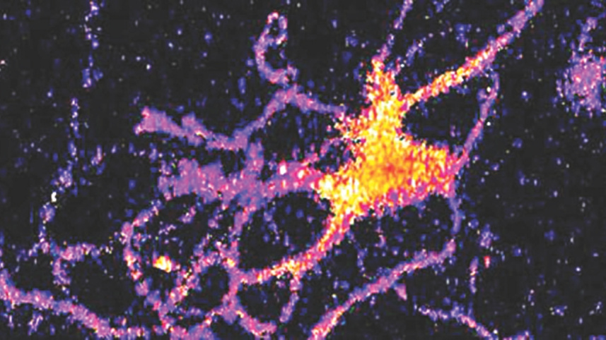
Brain Cells to Glow in the Dark
A new kind of bioluminescent sensor causes individual brain cells to imitate fireflies and glow in the dark.
The probe, which was developed by a team of Vanderbilt scientists, is a genetically modified form of luciferase, the enzyme that a number of other species including fireflies use to produce light. It is described in a paper published in the journal Nature Communications on Oct. 27.
The scientists created the technique as a new and improved method for tracking the interactions within large neural networks in the brain.
"For a long time neuroscientists relied on electrical techniques for recording the activity of neurons. These are very good at monitoring individual neurons but are limited to small numbers of neurons. The new wave is to use optical techniques to record the activity of hundreds of neurons at the same time," said Carl Johnson, Stevenson Professor of Biological Sciences, who headed the effort.
"Most of the efforts in optical recording use fluorescence, but this requires a strong external light source which can cause the tissue to heat up and can interfere with some biological processes, particularly those that are light sensitive," he said.
 For all latest news, follow The Daily Star's Google News channel.
Comments